Home

hendikepirani da li odbojka microscopia sem Spain Primirje Sažet Napušteno

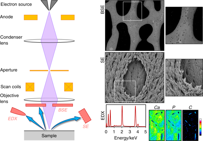
50 years of scanning electron microscopy of bone—a comprehensive overview  of the important discoveries made and insights gained into bone material  properties in health, disease, and taphonomy | Bone Research
50 years of scanning electron microscopy of bone—a comprehensive overview of the important discoveries made and insights gained into bone material properties in health, disease, and taphonomy | Bone Research

SEM-BSD image (left) and EDX spectrum (right): a Paper samples from an... |  Download Scientific Diagram
SEM-BSD image (left) and EDX spectrum (right): a Paper samples from an... | Download Scientific Diagram
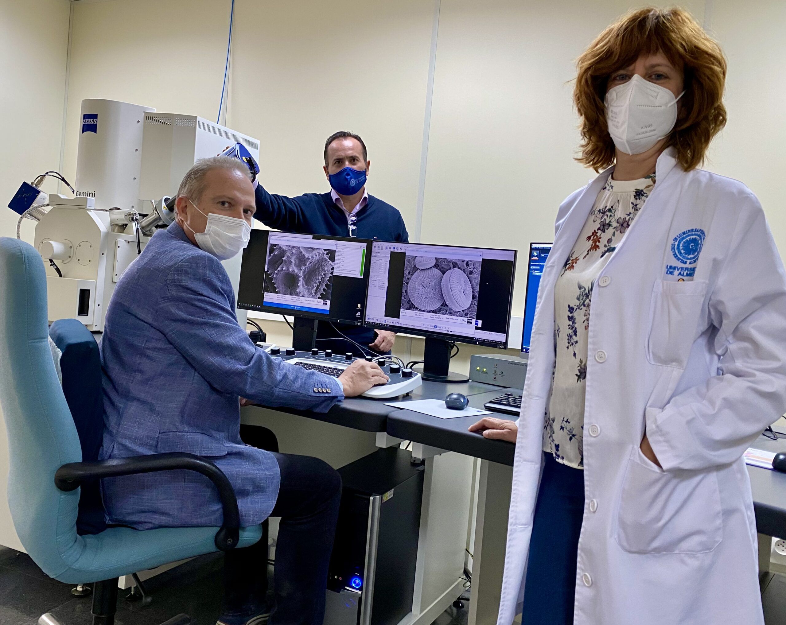
Nuevo Microscopio Electrónico de Barrido de Alta Resolución de Emisión de  Campo en la UAL | CDE Almería - Centro de Documentación Europea -  Universidad de Almería
Nuevo Microscopio Electrónico de Barrido de Alta Resolución de Emisión de Campo en la UAL | CDE Almería - Centro de Documentación Europea - Universidad de Almería

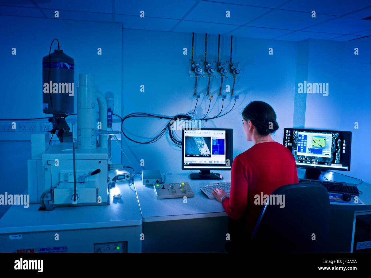
ESEM-Quanta | LMA
ESEM-Quanta | LMA

Analysis of microplastics - AIMPLAS

Microscopio electrónico de barrido de emisión de campo | Revestimientos  Rhenotherm No.1
Microscopio electrónico de barrido de emisión de campo | Revestimientos Rhenotherm No.1

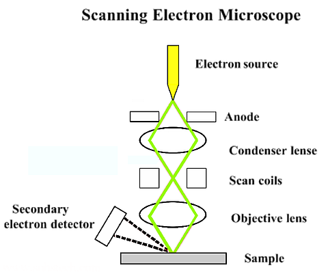
Scanning Electron Microscopy - SEM - Advancing Materials

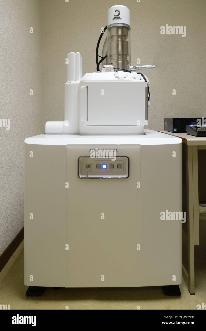
Scanning electron microscope atom hi-res stock photography and images -  Alamy
Scanning electron microscope atom hi-res stock photography and images - Alamy

SEM and LM images of stomata: a Sunken stomata for A. macrostachyum... |  Download Scientific Diagram
SEM and LM images of stomata: a Sunken stomata for A. macrostachyum... | Download Scientific Diagram
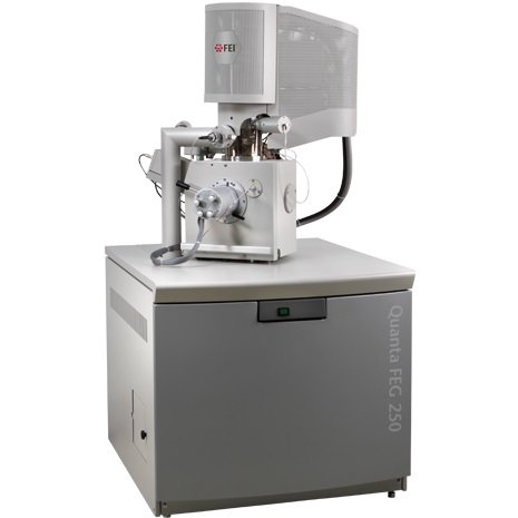
MICROSCOPIO ELECTRÓNICO DE BARRIDO AMBIENTAL SEM-QUANTA FEG-250, ESEM -  Elecmi
MICROSCOPIO ELECTRÓNICO DE BARRIDO AMBIENTAL SEM-QUANTA FEG-250, ESEM - Elecmi

Scanning Electron Microscopy Laboratory | Museo Nacional de Ciencias  Naturales
Scanning Electron Microscopy Laboratory | Museo Nacional de Ciencias Naturales

SME
SME

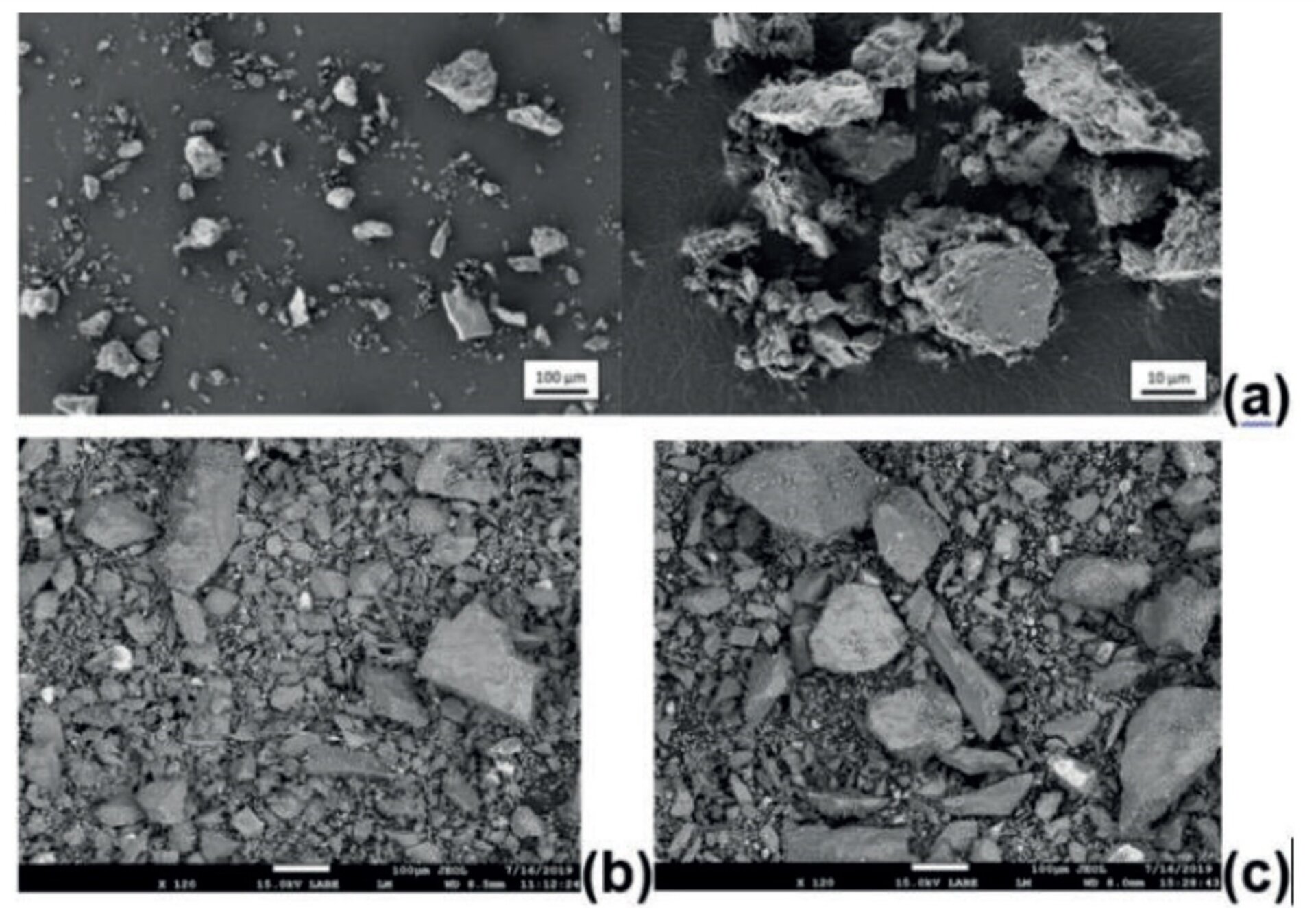
ESA - Microscopic close-ups of simulated lunar dust

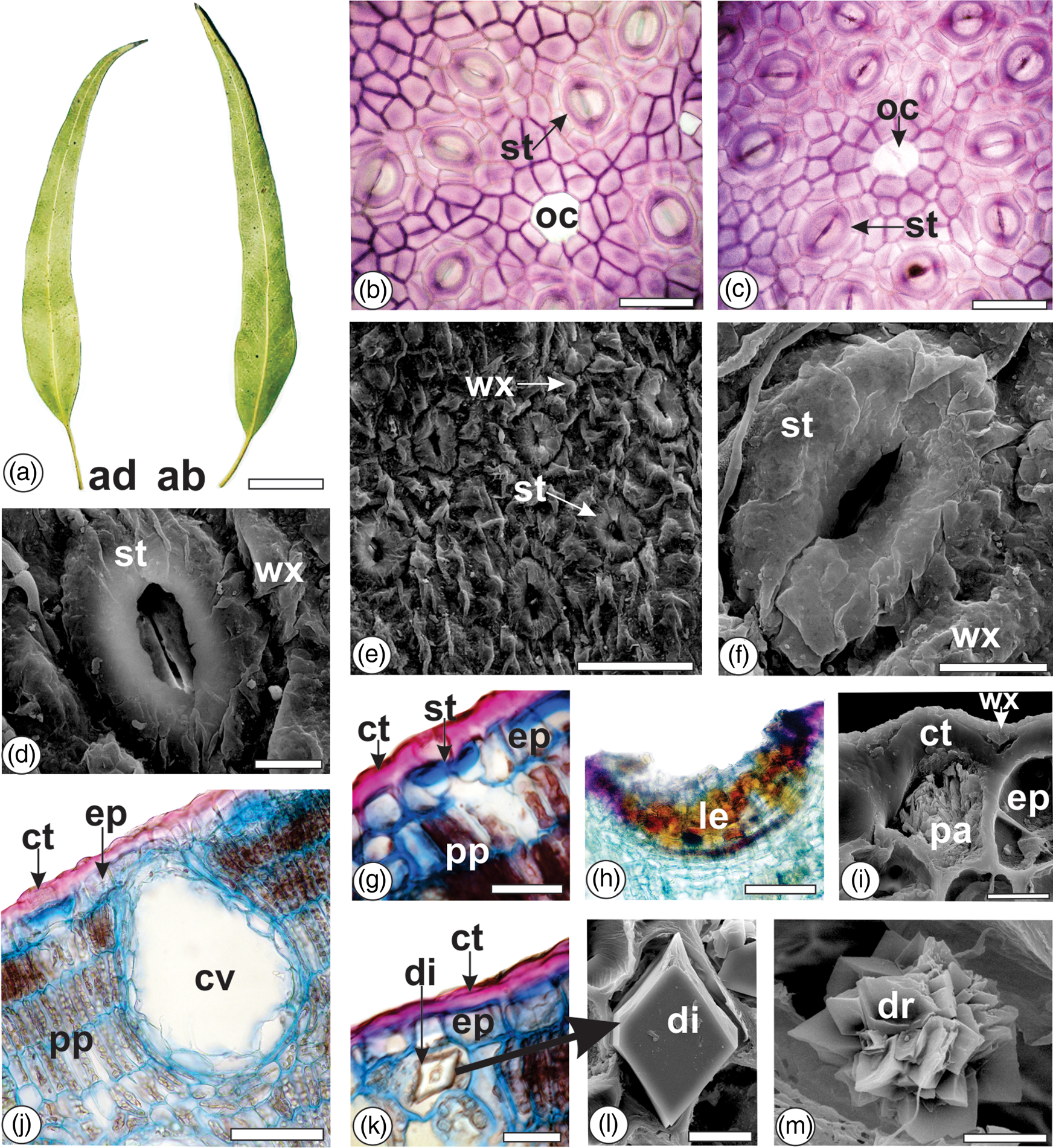
Light and Scanning Electron Microscopy, Energy Dispersive X-Ray  Spectroscopy, and Histochemistry of Eucalyptus tereticornis | Microscopy  and Microanalysis | Cambridge Core
Light and Scanning Electron Microscopy, Energy Dispersive X-Ray Spectroscopy, and Histochemistry of Eucalyptus tereticornis | Microscopy and Microanalysis | Cambridge Core
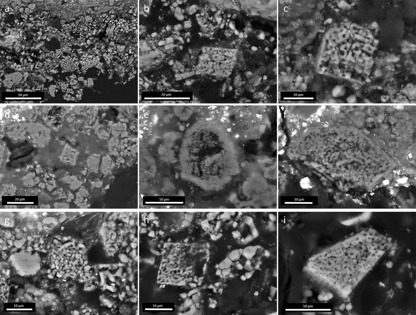
Plant Ash in Ground Preparations: Morphological Identification Uncovers  Novel Artistic Patterns in Baroque Paintings from Spain, North and South  America | The Metropolitan Museum of Art
Plant Ash in Ground Preparations: Morphological Identification Uncovers Novel Artistic Patterns in Baroque Paintings from Spain, North and South America | The Metropolitan Museum of Art

Correlative Voltammetric Microscopy: Structure–Activity Relationships in  the Microscopic Electrochemical Behavior of Screen Printed Carbon  Electrodes | ACS Sensors
Correlative Voltammetric Microscopy: Structure–Activity Relationships in the Microscopic Electrochemical Behavior of Screen Printed Carbon Electrodes | ACS Sensors

Biólogos Spain - La imagen de microscopia electrónica de barrido (SEM)  muestra glóbulos rojos y glóbulos blancos (células sanguíneas humanas) en  un pequeño vaso sanguíneo fracturado. La muestra se consigue mediante rápida
Biólogos Spain - La imagen de microscopia electrónica de barrido (SEM) muestra glóbulos rojos y glóbulos blancos (células sanguíneas humanas) en un pequeño vaso sanguíneo fracturado. La muestra se consigue mediante rápida

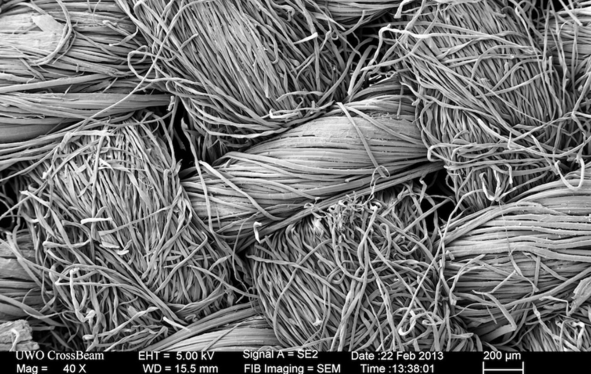
ESA - Microscopic view of textile

Biólogos Spain - La imagen de microscopía electrónica de barrido (SEM)  muestra una terminación nerviosa fracturada y queda expuesta parte de su  interior para revelar las vesículas sinápticas (naranja y azul) por
Biólogos Spain - La imagen de microscopía electrónica de barrido (SEM) muestra una terminación nerviosa fracturada y queda expuesta parte de su interior para revelar las vesículas sinápticas (naranja y azul) por

Scanning Electron Microscope - an overview | ScienceDirect Topics

Electron Microscopy - AnaPath

Biólogos Spain - Science Photo Library La microscopía electrónica de  barrido (SEM) nos ofrece una imagen coloreada de un ejemplar de la  mariposa, Ochlodes sylvanoides. Podemos apreciar la espiritrompa  (probóscide) y la
Biólogos Spain - Science Photo Library La microscopía electrónica de barrido (SEM) nos ofrece una imagen coloreada de un ejemplar de la mariposa, Ochlodes sylvanoides. Podemos apreciar la espiritrompa (probóscide) y la

Microscopio SEM - Nanos Sem

Adventures in Wood Microscopy, with Dr. Duncan Slater

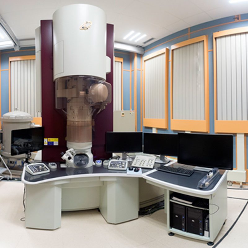
JEM ARM300CFEG: WITH CORRECTED ABERRATION IN OBJECTIVE LENS - Elecmi

Microscopio electrónico de exploración de KYKY Sem/instrumentación de  exploración de la microscopia electrónica
Microscopio electrónico de exploración de KYKY Sem/instrumentación de exploración de la microscopia electrónica

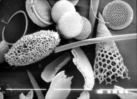
MICROFOSSILS
MICROFOSSILS

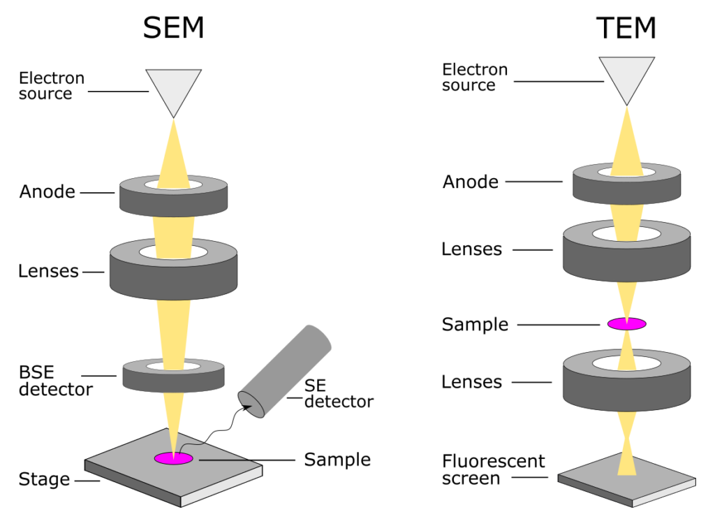
SEM vs TEM | Technology Networks

Scanning electron microscope hi-res stock photography and images - Alamy

Microscopía electrónica. Metrología óptica 2D y 3D.

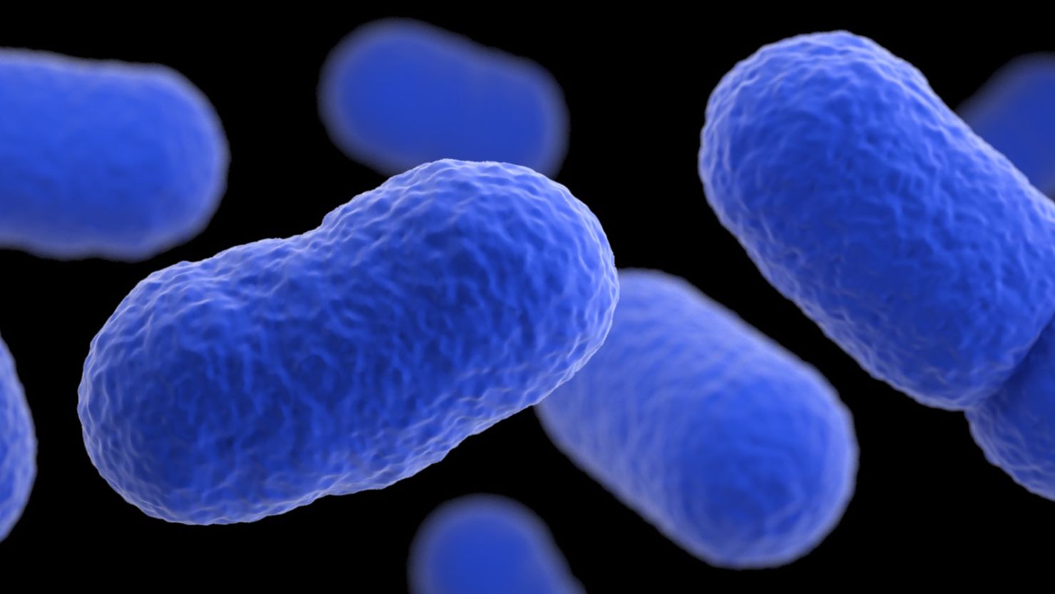
Spain issues listeria health warning as outbreak infects more than 170  people | CNN
Spain issues listeria health warning as outbreak infects more than 170 people | CNN

SME
SME

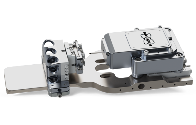
Hysitron PI 89 SEM PicoIndenter | Bruker

Fotografías de microscopía electrónica de barrido de semillas de... |  Download Scientific Diagram
Fotografías de microscopía electrónica de barrido de semillas de... | Download Scientific Diagram

Figure 3 from Electron Microscopic Study of the Illite-Smectite  Transformation in the Bentonites from Cerro Del Aguila (Toledo, Spain) |  Semantic Scholar
Figure 3 from Electron Microscopic Study of the Illite-Smectite Transformation in the Bentonites from Cerro Del Aguila (Toledo, Spain) | Semantic Scholar
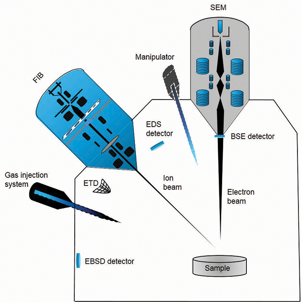
Focused ion beams: An overview of the technology and its capabilities -  2020 - Wiley Analytical Science
Focused ion beams: An overview of the technology and its capabilities - 2020 - Wiley Analytical Science
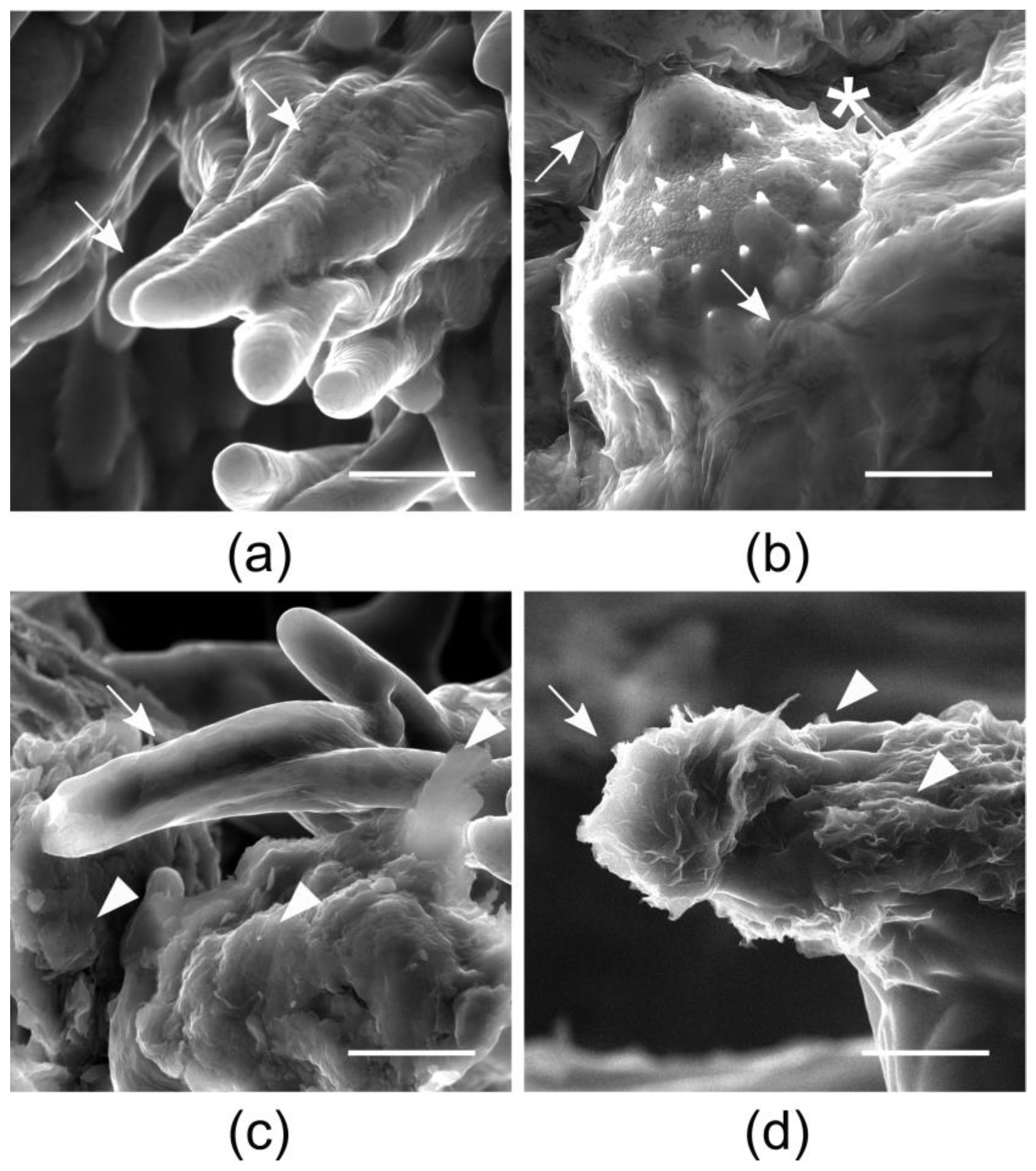
Applied Sciences | Free Full-Text | The Interaction of Graphene Oxide with  the Pollen−Stigma System: In Vivo Effects on the Sexual Reproduction of  Cucurbita pepo L.
Applied Sciences | Free Full-Text | The Interaction of Graphene Oxide with the Pollen−Stigma System: In Vivo Effects on the Sexual Reproduction of Cucurbita pepo L.
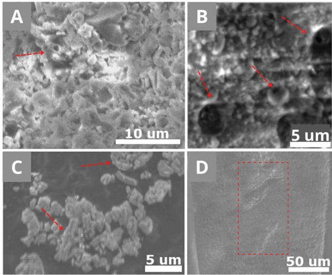
Conductive cross-section preparation of non-conductive painting  micro-samples for SEM analysis | Scientific Reports
Conductive cross-section preparation of non-conductive painting micro-samples for SEM analysis | Scientific Reports

Servei de Microscòpia i Difracció de Raigs X - UAB Barcelona

Scanning Electron Microscope Lab | Natural History Museum

CRYOELECTRON MICROSCOPY

Full article: SEM and light microscopic studies in seeds of Hibiscus  surattensis L. and phylogenetic attributes in Puducherry region, India
Full article: SEM and light microscopic studies in seeds of Hibiscus surattensis L. and phylogenetic attributes in Puducherry region, India

Seeds of Coronilla talaverae (Fabaceae), an endemic endangered species, in  Argaric Early Bronze Age levels of Punta de Gavilanes (Mazarrón, Spain) |  PalZ
Seeds of Coronilla talaverae (Fabaceae), an endemic endangered species, in Argaric Early Bronze Age levels of Punta de Gavilanes (Mazarrón, Spain) | PalZ

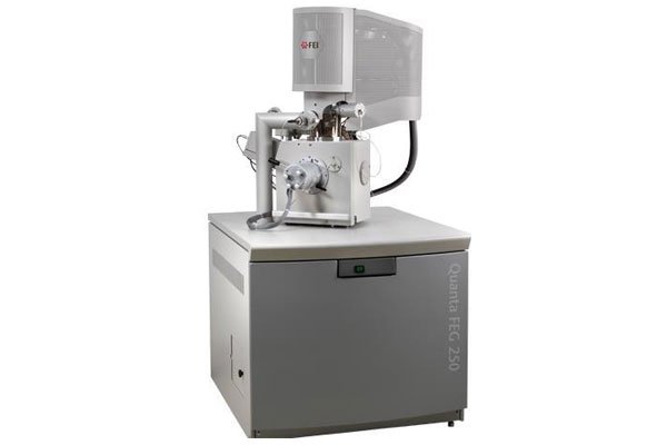
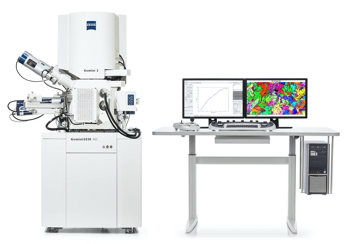
ZEISS introduces an integrated solution for multi-modal in situ experiments

Microscopía Electrónica de Barrido – SEM - Jasco Spain

ICMAB - Electron microscopy service

SEM images of coprolites of the hyaenid Lycyaena chaeretis (Gaudry,... |  Download Scientific Diagram
SEM images of coprolites of the hyaenid Lycyaena chaeretis (Gaudry,... | Download Scientific Diagram

CRYOELECTRON MICROSCOPY